Thursday, May 30, 2013

Final blog post

I really liked talking about diseases of the brain.  I learned a lot about Alzheimer's and strokes and tumors and I'd love to learn more.

Here are 2 links that my mom told me about a few years ago and I remember thinking these are cool pictures and I learned a lot so I suggest you check this out.

http://www.amenclinics.com/the-science/spect-gallery

http://www.amenclinics.com/the-science/spect-gallery/category/images-of-treatment

http://www.amenclinics.com/the-science/spect-gallery/category/images-of-treatment-2

here are 2 SPECT scans of an Alzheimer's patients brain


the blog post!!!

i liked the yummy neurons.
i learned allot from the owl pellets.


Aaron's last blog post

my favorite part of Anatomy class was when we did the owl pellets. and i feel like i learned a lot about the brain.                                                                                                    
thanks for reading.

Candy neurons!

I liked the candy neurons best this. They were tasty!
I learned a lot about the pig because we worked the longest on it.

Tuesday, May 28, 2013

NEURONS!!!

I really liked making the candy neurons and learned a lot making them as well! I think that letting kids eat the thing there learning about helps them remember it better, and that all teachers reading this should consider this blog post for future classes they might be teaching! But I leanrend a lot obout every thing coverd in this class!!!!!
THANK YOU JOLIEN!!!!!!!!!!!!!!!

Friday, May 24, 2013

Cranial Nerves -- 12 pairs of them!

This week, we focused on the 12 pairs of cranial nerves and what they do.


Here are some interesting things we noticed:
  • Sheep, cats, and humans all have the same 12 pairs of cranial nerves.  The size can be a bit different, like the olfactory nerves are proportionately larger in smell-oriented animals than humans, but the location and function of the nerves is pretty much the same.
  • Three of the 12 are motor nerves for the eye muscles.  They move the eyeballs in different directions.  I guess this must be very important.
  • Two of the 12 are involved in taste sensation: one for taste at the tip of the tongue and one for taste in the back of the tongue.
  • Some of them are sensory, which means they are transmitting sense information from the sense organ (eye, ear, nose, tongue) to the part of the brain that deals with that information.  Some of them are motor, which means they are transmitting a signal from the brain to a muscle to tell that muscle to move.  Some of the nerves are bundles of axons that have a sensory part and a motor part, so they are considered both sensory and motor nerves.
  • The numbering of the cranial nerves is in roman numerals.  They are numbered I through XII (1 - 12) starting from the front of the brain and moving towards the back of the brain (looking down at the underside of the brain).
  • The names of the nerves are:  
    • I: Olfactory: smell information going from nose to brain
    • II: Optic: visual information going from eyes to brain
    • III: Oculomotor: moves your eyeball and constricts pupil
    • IV: Trochlear: moves your eyeball (downward and in towards your nose)
    • V: Trigeminal: senses touch from the face and head, and moves your jaw for chewing
    • VI: Abducens: moves your eyeball (side to side)
    • VII: Facial: taste information going from front of tongue to brain, muscles of facial expressions
    • VIII: Vestibulocochlear: hearing and balance information going from ear to brain
    • IX: Glossopharyngeal: taste information from back of tongue, and swallowing movement
    • X: Vagus: sense and movement in your glands and digestive system
    • XI: (Spinal) Accessory: controls muscles for head movement
    • XII: Hypoglossal: controls tongue muscles
  • There are lots of mnemonics for the cranial nerves.  A mnemonic is something you use to remember something (often a list of things).  Typically, you make a phrase that you can remember that uses the first letter of each item on the list.  For the cranial nerves, here are some of the mnemonics I found:
    • Odor OOrangutan Terrified Tarzan After Forty Voracious Gorillas Viciously Attacked Him
    • OOccasion Our Trusty Truck Acts Funny, Very Good Vehicle Any How
    • Once One Openly Told Tourists About Fighting Vampires Gobling Various Antelope Herds
    • Only Owls Observe Them Traveling And Finding Voldemort Guarding Very Secret Horcruxes
    • OlOpie Occasionally Tries Trigonometry And Feels Very Gloomy, VaguAnd Hypoactive
    • OlOprah Occasionally Trots Triumphantly About, Farting Velveeta Globs, Vaunting Accolades Hysterically
    • Once OOctober Thirteenth, Troublesome Abductors Filched Various Golden Valuables And Heirlooms
    •  Old Oppressive Oceanic Trout Trick Aquatic Fauna Very Greedily; Valiant Sharks Hunt (them).
    •  Oh Once One Takes The Anatomy Final, Very Good Vacations Are Heavenly.
    • A visual mnemonic for the cranial nerves!
    • Here's one I just made up: Often, one opens the top and front velcro, giving villains a hand.
  • There are also mnemonics to help you remember which ones are sensory (S), which are motor (M), and which are both (B). 
    • Some Say Marry Money, But MBrother Says Big Brains Matter Most
    • Silly Superman Made Mortal Brothers Make Bets Since Both Boys Made Money
    • Some Say Money Matters, But MBeloved Says Being Beloved Matters More
    • Here's one I just made up now:  Share Some Merry Music By Making Brother Shake Both Bongos Mighty Madly
Testing the Cranial Nerves

In class, we did activities to illustrate what daily activities use which cranial nerves.
For the Olfactory Nerve (Cranial Nerve I), we had four boxes that each contained a cotton ball with a scented liquid on it.  We each sniffed the cotton ball, then guessed what the smell was.  The four scents were:
  1. vanilla (we discussed why it smells like alcohol -- because the vanilla beans don't dissolve in water, so alcohol is used to get the flavor into a useful form)
  2. orange extract
  3. vinegar
  4. lemon extract
For the Optic Nerve (Cranial Nerve II) we took an eye test.  Students in the class have excellent eyesight!

For the Oculomotor, Trochlear, and Abducens Nerves (Cranial Nerves III, IV, and VI) we moved our eyes in all directions, tracking the movements of my finger without moving the head.

For the Trigeminal Nerve (Cranial Nerve V), we ate some food -- using the muscles that move our jaws to chew -- and I also touched each student's cheeks with a cotton ball.  

Making faces -- cranial nerve VII
For the Facial Nerve (Cranial Nerve VII), we made funny faces and also tasted brownies with the front parts of our tongues.

For the Vestibulocochlear Nerve (Cranial Nerve VIII), we tested our hearing by closing our eyes and raising hands when we hear a quiet snap across the room.  Excellent hearing in all the students.  Then, we balanced on one foot and closed our eyes.  No one fell down!

For the Glossopharyngeal Nerve (Cranial Nerve IX), we swallowed a sip of water.

We forgot to do anything with the Vagus Nerve (Cranial Nerve X) because the website we looked at didn't show anything.  But of course we were using our Vagus Nerve as we digested the foods we were eating!  Students brought bloody rice krispie treats, eyeball mozzarella balls, and breadstick fingers with almond fingernails.  Brownies and carrots were there to balance it out.

For the Spinal Accessory Nerve (Cranial Nerve XI), we partnered up and had one person put their hands on the side of the other's head.  The partner then tried to move their head side to side.  

For the Hypoglossal Nerve (Cranial Nerve XII), we all stuck out our tongues, moved them to the right, then to the left, then back into our mouths.

We all passed with flying colors, and appear to have healthy cranial nerves!!

Wednesday, May 22, 2013

Strawberrys

Has any body else noticed how strawberry's sort of look like brains?
They have wight "matter" in the middle and red "matter" on the outside and in the very middle, see? Maybe there structure is designed sort of the same.


Sunday, May 19, 2013

Nervous System Diseases and Their Anatomy

This week we focused on diseases and disorders of the nervous system.  One lead-in to this unit was our homework assignment, which was to do an online virtual brain surgery for treating Parkinson's Disease.  All of us who did the simulation enjoyed it a lot.  Basically, the operation involves the implantation of electrodes into the brain areas that are damaged in Parkinson's Disease.  As I explained to the students, the neurons in that area die and never grow back.  We don't understand exactly how those neurons function, but stimulating that area electrically is very helpful for reducing and sometimes eliminating many of the symptoms of the disease.

During the game, you first see the patient with her symptoms, which are that her hands shake (the "tremor" that is typical of this disease).  Next, you go through all the steps of the procedure: inserting screws called fiducials into the skull, using them to precisely map the path for the electrodes to follow, drilling into the skull and placing the equipment for guiding the electrodes, slowly and carefully pushing the electrodes into position, waking the patient to use their reported sensations to show whether they have been placed correctly, closing up and removing the equipment, then two weeks later testing for the correct power, placing the batteries into the chest region and connecting them to the electrodes.  Another more complete explanation is here.  It was detailed and fascinating to actually do the simulated surgeries!

After discussing the simulation and why we did it, we moved on to an active discussion of nervous system diseases and disorders.  There are a huge number of problems that happen with the nervous system, and I fully admitted that I am not an expert on all of them, or even very many of them.  I have studied a few nervous system diseases in depth, which we discussed, but most of what we are talking about are diseases that I know a fair number of basic details about.

I brought in two brain models that I borrowed from the UW Neuroscience Training Program.  One was a rather large, colorful brain model labeled with each region's function.  We didn't spend a lot of time on it, but it is very helpful for seeing exactly where the regions we are discussing are located.  It also illustrates the mapping of the body parts onto the motor area (where the neurons are that tell that body part to move) and the sensory area (where the neurons are that tell us we are sensing touch at that body part).  It comes apart into four parts, so you can see some inside brain regions too, but we couldn't find the substantia nigra (area first affected in Parkinson's Disease).

The other model was specifically designed to illustrate a number of fairly common nervous system diseases.  Unfortunately, it was missing the informational card that would confirm for sure which parts of the model were supposed to illustrate what, and also there was a piece of brain blood vessels that was missing.  The diseases modeled were: 
  • alcoholism
  • Alzheimer’s disease
  • aneurysm
  • depression related tumor
  • seizure related tumor
  • migraine
  • multiple sclerosis
  • Parkinson’s disease
  • stroke
  • subdural hematoma

We only had time to discuss some of these diseases.  For each disease, we got up out of our seats and made a respectful effort to act out the symptoms of the disease.  This was to solidify what we discussed, get a feel for what it might be like to suffer from the disease, and of course to stay active.  Here's what we actually discussed:
  • Parkinson's diease begins with the loss of neurons in the substantia nigra, and the model showed us two substantia nigras:  one looks like a little, blackish, half mustache, and the other was blank.  The blank one had lost over 90% of the neurons that had been there, and showed what that area looks like in a Parkinson's disease patient.  Normally, there are I think at least a million neurons in the substantia nigra on each side.
  • Alzheimer's disease is a progressive disease that first and most deeply affects the memory.  It is a disease that involves dementia, or a specific type of memory loss.  Dementia is a general term for the type of memory loss in which the patient has severe enough problems with thinking, memory, and reasoning that is severe enough to interfere with daily functioning.  Sometimes the person will forget people they've known for a long time or forget what they're doing.  
    • I once cared for a woman with advanced Alzheimer's disease for an evening, and it was very interesting and sad to see firsthand the effects of the disease.  This woman told the same brief story over and over again, seeming not to know that she had told the same story just a few minutes before.  She had been retired for years, but forgot that and had a hard time believing me when I told her that she didn't need to go to work.  Her apartment door needed to be locked to prevent her from wandering out and getting lost.  
    • As you can see in the picture to the right, many areas of the brain get much smaller in advanced Alzheimer's disease.  Vision and motor areas are not badly affected, and patients don't have a lot of problems with movement or sight, but memory areas are destroyed, olfactory areas (sense of smell) are damaged, and the frontal lobe of the brain, which helps us think ahead and plan, is severely affected.
  • Aneurysm is when a blood vessel in the brain has a weak spot that then fills up like a balloon.  Eventually, it can leak and/or rupture, and this is a major problem.  Doctors can do surgery to fix the area by clipping the aneurysm.  If they do this in time, the patient is fine, but if the aneurysm ruptures, there is generally no way to save the patient and he or she dies.
    • A friend of my had an aneurysm that started to leak and might have ruptured if it hadn't been
      caught in time.  Her symptom was a severe headache like she never felt before, and luckily she was right across the street from Duke University Medical Center when it happened (one of the best hospitals in the country, #8 on US News & World Report).  They gave her top notch care, including a relatively new procedure involving raising the patient's blood pressure to prevent the brains blood vessels from closing.  When this happened, I immediately did research to better understand aneurysm and learned that if the patient survives the aneurysm by getting it surgically fixed before it ruptures, blood that leaks from the aneurysm causes a reaction over the next few weeks in which blood vessels can close up.  If that happens, it is almost like a stroke because that part of the brain stops getting blood and oxygen, which can cause permanent damage.  Fortunately, my friend was able to fully recover with no permanent damage, which was at least partly due to excellent care at Duke!
  • Stroke is when a blood clot gets stuck in the brain and causes a small or large part of the brain to be without blood and oxygen for a period of time.  There are blood vessels all over the brain, and where the clot gets stuck determines what part of the brain is damaged and therefore what the patient's
    symptoms are.  If the speech area is affected, the person may not be able to talk.  If the area relates to memory, they may have memory loss.  Often many areas are affected, and the person may have some paralysis, some speech problems, and some memory loss.  
    • There's an interesting book called My Stroke of Insight, which is written by a neuroscientist who had a major stroke. She survived and recovered, and was able to describe what it was like.  It was so severe that she lost her understanding of what numbers were and what a phone was for, and so even though she was near a phone, she couldn't figure out how to call someone for help!  Luckily, someone called her, and hearing her speech get weird, they knew she needed help and got her to the hospital right away.
  • Migraine is a special type of headache that is very severe and is thought to be related to dilation of blood vessels in and around the brain.  For some people, there is an aura, which is usually a visual phenomenon like flashes of light a few minutes before the migraine hits.  
  • Brain tumors are cancer cells in the brain.  Cancer cells grow and divide without properly obeying the normal signals to stop growing.  Some brain tumors invade healthy tissue and damage it, while some stay separate from the healthy tissue but push into it and cause brain damage due to the pressure. 

Thursday, May 16, 2013

amyotrophic lateral sclerosis or ALS or Lou Gehrig's disease

Amyotrophic lateral sclerosis is a brain disease that affects the part of your brain, brain stem, and spinal cord that controls muscles we control by thinking about, and don't move by them selves (like your heart does). You have two sections of neurons that mainly do this, the upper and the lower. The upper is in your brain and the lower is in your spinal cord. the neurons in these parts of brain, spinal cord, and brain stem are called "Motor neurons". The disease is were the motor neurons slowly decrease in function and eventually stop sending messages to muscles like the ones that control your arms and legs. The victims amyotrophic lateral sclerosis also lose function in there muscles in the diaphragm and chest resulting in failure to breathe without some kind of respiratory system. if you want to know ALS is sometimes called Lou Gehrig's disease, read this artical, Its about a baseball player named Lou Gehrig who had ALS. ALS is fatal, and most patients die within 4-5 years. But some (about 10%)  live for 10 years.
If you want to know more about ALS go to this site.

Wednesday, May 15, 2013

Touching a human brain

This week, we each got to touch a real human brain.  The Neuroscience Training Program at the University of Wisconsin Madison has an outreach kit that they allowed me to check out for demonstration for the students.  Included was:


  • one whole human brain, fixed, with the membranes on it.  No information was available on the person who donated it, for confidentiality reasons.  So all we know was that the person passed away and had chosen before their death to donate their brain to science.  It was pretty large, so we thought perhaps it belonged to a male.
  • one half human brain, fixed.  Because it was cut down the middle, we could see the inside parts of the brain easily.  I'll detail those below.  It was smaller than the whole brain, so perhaps it belonged to a woman.  
  • gloves and a tray for looking at the brains.
  • two brain exhibits, showing the brains with eyes of several animals.  One had perch, frog, snake, and cat, while the other had 
  • a bag of foam brains, like the one shown here:  --->  They are squishy, like stress balls, and we could each have one to take home.  We used them later on, in one of our games.
  • two mirror writing set-ups, which are boxes that show how we use our brains to learn new skills.  You put the box together and then put a paper in the bottom, which you can see through the mirror but not directly.  You have to draw between the lines of the shape by looking through the mirror.

Neuroanatomy & The Sheep Brain Dissection

This week, we really got into the brain anatomy more deeply and did the sheep brain dissection.  First, we discussed human brain anatomy and watched this movie.  It goes through some basic brain anatomy and uses a 3D model system to show different parts.  We also discussed a variety of brain topics, including watching this video of a girl who had half of her brain removed due to severe epilepsy.  During the video, I went over these details:

  • The right side of the brain controls the left side of the body, and the left side of the brain controls the right side of the body
  • So, her paralysis on the left side of her body shows that it was the right side of her brain that they removed in the surgery.  
  • It's pretty amazing that the remaining half of her brain was able to take over a lot of the work of the missing half, so that she can walk and talk, and the droopiness of the left side of her face became much less noticeable.  She was able to re-learn most of what she needed to and is doing well in school
  • They did not remove some of the very deep structures of the right side of her brain, because they are important for basic things like breathing and keeping the heart beating.
We talked about another brain disease before the dissection, which was Parkinson's Disease.  I showed the students what part of the brain is affected when you have PD, which a small area called the substantia nigra.  The reason it is called that is because it is one of the only naturally pigmented areas of the brain, and it looks like little iron filings scattered in a mustache shape deep in the brain.  People don't have any symptoms until about 90% of the neurons there have died.  They first notice a little twitch, like in their pinky finger, and then they gradually develop a tremor.  The tremor can be treated for a while with various drugs that make it go away for a few hours, but then it keeps coming back and getting worse and worse.  Eventually, the person has more and more problems and passes away, typically after 20 or more years of symptoms.  For the homework, we will explore a surgical technique to help with PD symptoms.
The above image is a view of the brain by a coronal section, as illustrated in the small figure.  We talked about cutting a brain horizontally and sagitally too.  Here is a picture of the different ways of cutting the brain.  During most of my research, I have done sagittal sections of brain, and that is what we did for the sheep brain dissection.  To do a sagittal section, we cut the brain down the middle from above.  


For the dissection itself, we followed the student guides and also this procedure.  Some things we talked about during the dissection:
A view of the dissected sheep brain

  • Meningitis is when the membrane covering the brain (called the meninges) gets infected.  It is very serious and can kill you, because swelling within the skull causes high pressure, which then squishes the brain and causes irreversible damage.
  • The bulges all over the surface of the brain are called gyri (plural of gyrus), and the grooves are called sulci (plural of sulcus).
  • There are a lot of nuclei in the brain (plural of nucleus), which are groups of neurons that do some specific function.
  • The hypothalamus is a small structure near the brainstem that has several nuclei.  One of them controls things like hunger, thirst, and sleep.
  • The pituitary is a gland that releases hormones, including growth hormone to tell the body when it should grow.
  • Sheep have large olfactory bulbs that they use for understanding smells in their world.  Most mammals rely more on smell than we humans do, and their brain olfactory areas are proportionately larger.
  • The cerebellum is important for learning motor skills, like riding a bike, mirror writing, and throwing a ball.
    • My neuroscience program has several sets of prism glasse
      s they use for demonstrations.  You throw a ball into a basket very easily, then put on the glasses.  Now, you start missing the basket.  If you keep trying, your cerebellum helps make the right adjustments to your throwing and you start to get baskets again.  Then when you take them off, you start missing again and need to keep practicing to adjust back to how to throw.  This all happens within seconds to minutes!  Learning to ride a bike takes a bit longer, but once you get the hang of it, you don't use your cerebellum much for that skill anymore.
    • The ventricles of the human brain
    • A man who had a major memory impairment due to stroke lost his ability to make new conscious memories.  Everyone he met after his stroke he promptly forgot who they were.  But he worked with researchers over many years who taught him motor skills like mirror writing.  While he had no memory of ever trying the task, his skill improved at it, showing that the motor learning happened in a different part of the brain than his stroke damaged area.
  • The corpus callosum is where axons from neurons in the brain cross over from one side to the other.  Surprisingly, people function pretty well if their corpus callosum is cut or never forms.
  • Ventricles in the brain are areas with fluid in them instead of cells.  The fluid that the brain and spinal cord sit in is called CSF, or cerebrospinal fluid, and it helps keep the brain healthy.


Thursday, May 2, 2013

Brain Video

Hey my mom showed me and my brother this video and thought it might be good for tomorrow sorry for doing the homework late


Hope u guys get a kick out of it like we did

Monday, April 29, 2013

Yummy Neurons!

This week, we made models of neurons -- out of candy!  It was a team effort, and it seemed like everyone was able to leave with a sugar high!  There was enough candy for all the parts of interest, and we even had some choices to make:

  • The cell bodies were the cupcakes.  Some cut off the tops and dug out the middle, and some just dug into the top of the cupcake to create space for the insides of our neurons.
  • Once we had space, in went the cytoplasm -- green Jell-O!
  • We each chose a nucleus for our cells -- either a large jelly bean or a cherry sour
  • Before putting the nucleus into the cytoplasm, we wrapped bits of endoplasmic reticulum around them -- fruit roll-ups
  • On some parts of our endoplasmic reticulum, we sprinkled some ribosomes, which makes it rough endoplasmic reticulum (smooth endoplasmic reticulum doesn't have ribosomes).  Most of us used red sprinkles for the ribosomes, but we discussed using nerds.  Some sprinkled nerds into the cytoplasm to represent the ribosomes that float loosely separate from the endoplasmic reticulum.
    Someone else's candy neurons; you can see organelles!
  • Next, we chose some mitochondria -- small jelly beans, about 3 per cell (most wanted the Starburst jelly beans, but a few used the Jelly Bellies)
  • Some of us added lysosomes to our cytoplasm -- nerds
  • One student who just got his braces put on used raisins for his organelles, since he couldn't have some of the gooier candies
  • Then, we added the key parts of neurons that make them special compared to other cells:
    • The axon was a Twizzler, and we wrapped it with myelin sheaths -- marshmallows
    • The dendrites were bits of sour licorice straws
    • We added tiny synaptic boutons, which are where the signal comes into the dendrites
    • To represent the neurotransmitter crossing the synapse to send the message from one cell to the next, we used the frosting
Finally, we created a large circuit with our neurons, by connecting them together.  We lined up our neurons and connected each axon to one of the next cell's dendrites (with neurotransmitter of course), and created a seven or eight cell circuit!  Most of our neurons did not survive long after that, moving bit by bit into our digestive systems.
Diagram of two neurons showing their synapse

To model how neuronal circuits work, we also lined up and held hands, to represent neurons connected together.  A message started at one end of the circuit and traveled to the other end.  The message was a hand squeeze, and each neuron had to send the message on to the next as soon as it was felt.  The last neuron said "Got it" when the message got through.  We all noticed that it was a bit slow the first time, but after repeating the process several times, the message passed very quickly.  Just like when the neurons get myelinated, so their messages travel much faster.

We send some more messages down our circuit using the game "Telephone" where one person whispers a message to the next person, who whispers it to the next.  One message sent that way arrived intact, but several others were changed or garbled.  The nervous system has many features to prevent that kind of thing from happening.  Its job is to send messages very quickly and very accurately all over our bodies.

Monday, April 15, 2013

Neuroscience intro

We are finally at my area of expertise -- neuroscience -- and we started by reading this page for homework and having a quiz show based on it.  Here were some of the main facts we went over:

  1. Brain cells or nerve cells are called neurons.
  2. Neurons have a nucleus, cytoplasm, mitochondria, and other organelles that are typical of animal cells.
  3. Neurons communicate with each other.
  4. The way they connect and communicate is through structures called synapses.
  5. Synapses are places where neurons communicate, but there is a teeny tiny gap between the two cells.
  6. The brain has approximately 100 billion neurons, and approximately 1 quadrillion synapses.
  7. The adult human brain weighs about 3 pounds
  8. Neurons have a long, skinny part called the axon that sends the message to the next cell.
  9. If you laid all of an adult human's neurons end to end, it would be about 600 miles.
  10. The brain is about 2% of the total body weight for an adult.
  11. Most neuroscientists have had 12 years of grade school, 4 years of college, and 4-7 years of graduate school.
We also discussed the insulation around neurons.  You can see in the image above that there are rod-like things going down the axon. That is actually from cells called oligodendrocytes that are near neurons that wrap themselves around the neurons to insulate them, like we use rubber to insulate wires.  The insulation makes the message travel faster, and as our brains get more insulation around their neurons, we become more able to do things.  Babies have hardly any of it, and new skills develop as their brains
develop in this way.  If you lose the insulation around your neurons, you become disabled in various ways because the brain doesn't function as well.
The insulation is very fatty, and because of that, areas of the brain and spinal cord with a lot of insulation look white.  Areas that mostly have the cell bodies of the neurons look gray.  That's why these areas are called white matter and gray matter -- they really look white and gray when you dissect them, as we will see.

We took a look at our sheep brains and saw that they have a thin
What our sheep brains look like
membrane over them.  This is not much real protection compared to the skull, but it helps regulate the fluid pressure inside the brain, which is very important.  Too much or too little pressure inside the brain can be a disaster!  We also saw the brainstem at the base of the brain, which is a huge bundle of mostly axons that are going to the spinal cord.  We noticed that the surface feels smooth, but you can see all sorts of wrinkles and twists like a small intestine.  We will discuss why that is and what it means.  Lastly, we weighed our brains, and they weighed about 150g.  Not very much!  They are very small compared with human brains!

Thursday, April 11, 2013

Awesome cell stuff!

In class i learned that the nucleus holds the hole blueprint to make a hole new cell!
And that the cell wall holds up the side of the plant cells!
It was really fun to look at the dyed slide!


A dyed slide!     

Monday, April 8, 2013

Slide-Making and Cells

This blog post covers what we've done for the past two class meetings.  Our overall focus for these two classes has been making slides, and for the second class, we discussed what cells are and the parts of cells.

Histology is the microscopic study of the cells and tissues of plants and animals.  Basically, that means that people put samples onto microscope slides and the look at them under the microscope to see what they can find out about the sample.  Most of the time, they add a stain to the sample, because you can't see very much without a stain.  Stains are usually dyes, and they are added to the sample because they often stick more to some parts of the sample than others, which lets you know things like where the edges of the cells are and where parts within the cells are.

Here is a picture of kidney tissue stained with H&E, which makes some parts of the cells pink, some red, and some dark purple.  It's a very common stain for human or animal tissues.
At some point, we would like to stain the tissues we are examining from our pigs with eosin dye, but it might not work very well for several reasons.  First, there is a complex staining process that will be hard for us to replicate, and second, we don't have the equipment to cut our samples super thin.  Most histology samples are about 4 microns thick, cut using a microtome.  A microtome works basically like a meat slicer and cuts very thin sections of your sample, that you then put onto a slide, stain, and examine under a microscope.  I will look into the possibility of us getting a tour of a histology facility on campus, as there are several decent ones in the medical school and vet school.

During the first class, we took samples of our own cheek cells and looked at them under a microscope.  Here's what they looked like without any stain on them and with methylene blue stain on them:
You can see that the blue stain lets us see the cells better, including the little oval in the center, which is the nucleus.


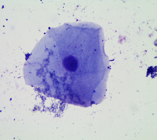
We also tried a purple stain called crystal violet, and cheek cells stained with it look like this: 

I got this image from the internet, and I think the stain makes bacteria dark purple, which would be the little tiny dots all over the place.  We have a lot of bacteria in our mouths, and this picture shows how much more bacteria we have than even our own skin cells.

We tried Eosin too and didn't see much, but also our microscope is dying.  The update is that the nosepiece, which is the part that has different magnification objectives, came off.  I don't think I can fix it, so I think we have to let that really cool microscope rest in peace.  I still have the dissecting microscope, and will look into getting another high magnification microscope.  

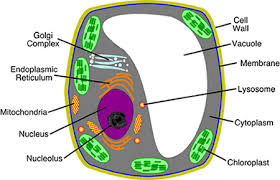
This past week, we discussed the parts of plant and animal cells, so that we can better understand what we're seeing when we look at cells under a microscope.  We assigned each student a cell part to represent, and discussed what each part does.  

First, Henry was the cell wall.  The cell wall is only found in plant cells, and it is a rigid structure that helps the plant stay upright, grow tall, and keep the cell protected.  Animal cells don't have one because they need to be more flexible, such as muscle cells that need to contract.

Then, Aaron was cell membrane.  Animal cells have a cell membrane to separate them from their environment, and so do plant cells.  It is more flexible than the cell wall.

Elinor was the nucleus, which is a very important organelle.  (Organelles are the small parts inside cells that do things for the cell, like organs in our bodies do things for our entire body).  The nucleus contains the DNA, which is the complete blueprint for making the whole organism.  It's like a recipe book, and each recipe makes a protein.  If you follow all the recipes in the right order, you make an Elinor or a frog, whatever the blueprint (DNA) says.  Every cell in our bodies contains a complete blueprint for making us, with two exceptions.  Red blood cells are born with nuclei (plural of nucleus), but after a few weeks, once they have made the proteins they will need to carry oxygen around the body, they spit out their nuclei and live for another month with no nucleus.  Sex cells (the sperm and the egg) each only have half of the blueprint, such that when they combine, a complete blueprint for a person is made.  If each had a complete set of DNA, there would be too much and an organism can't be made if a cell has a double blueprint (too much DNA).

Jett was the vacuole, which is the large white space we can see in the cells.  In plant cells, there is a large, central vacuole that takes up most of the space in the cell, but in animal cells, if they even have vacuoles, they are small and don't take up much space.  It's not clear to me exactly what they do.  

Anne Sophie was the endoplasmic reticulum, which will always be found around the nucleus.  It is a stack of tubes that take information from the DNA blueprint to turn into proteins.

Esme was the lysosome, which is a small blob in which proteins that are no longer needed get broken down.

Ben was the mitochondria, which makes the energy for the cell.  Plants have chloroplasts in some of their cells, which take energy from the sun to start making sugar.  All cells have mitochondria that take sugars from photosynthesis or from eating foods and then convert them into energy the cell can use to make things and live.

Our plant cell
Jordan was the cytoplasm.  All the organelles float in a liquid called the cytoplasm.

Liam was the ribosomes, and some ribosomes just float around on their own while many of them are actually in the endoplasmic reticulum.

There are more organelles than we discussed, but we're going to leave it at that for now.

Next, we pulled off the skin layer of some onion bulbs and looked at them under the microscope.  Here are some pictures we took.
High magnification view of our onion cells
Unstained by slightly dried out onion skin

Onion cells stained with Iodine-propidium iodide
 
Lastly, we looked at a section of lilac leaf that was stained to show us the different cells.


Monday, March 18, 2013

Finally -- Done with the Pig Dissection!

This week, we finished our pig dissections.  Some of the students were interested doing the final step of sewing up the Y incisions we made, like what is done for autopsies.  The organs that have been removed and examined are placed back in the body, arranged to restore the natural shape of the body.  We did this with our pigs and used suture to sew them up.

Before that, we had our quizshow about the kidneys, and the students who didn't want to sew up the pigs watched a series of educational videos related to anatomy (from a YouTube playlist I made).  At the end of the playlist were some Schoolhouse Rock videos, which I remember from my childhood.  I decided to play two additional YouTube videos that illustrate the organism classification system.

This one is from 1989, and is the first video putting the system to a rap beat.  Just to remind you, here are the groupings, from most general to most specific:
Kingdom
Phylum
Class
Order
Family
Genus
Species

Most organisms that you can find on Wikipedia have their groupings listed with their entry.
Here's the second classification rap video we watched:

We have common names for organisms, such as fruit fly, and we have their scientific name, which is their Genus (capitalized) and species (lowercase).  Fruit flies' scientific name is Drosophila melanogaster.

Next week, we will be making slides of our tissues and staining them to look at under the microscope.

Thursday, March 14, 2013

LHK

Why is the liver so important it filters stuff out of your blood like alcohol

how does the heart pump blood   electrical impulses sent down by "nodes" thru conducting pathways towards the atria and ventricles.

what does the kidney do it produces and generates urine


Quiz

1:Why is the liver so important?
2:How does the hart pump blood?
3:Why does the kidney do?

Wednesday, March 13, 2013

Kidneys -- They're not just cool beans!

This week's focus was the kidney, an important organ whose main role is to filter the blood to generate urine.  We can live without one kidney, but not without two.  They are located in the abdominal cavity near the organs of the digestive tract, but they are part of the urinary system, not the digestive system.

This is the machine for dialysis
The process of breaking down food to get the nutrients leads to waste products, including urea.  If these waste products are not cleaned out of the blood, we will die.  When someone's kidneys are not functioning well, he or she must go on dialysis to survive.  Dialysis involves being hooked up to a machine that takes out some of your blood, cleans and filters it, then puts back the filtered blood.  It is not as good as your kidneys, but if you go for dialysis often enough (typically at least 3 times a week for several hours), you can survive for a long time without functioning kidneys.

Kidneys make sure that sugar does not get into the urine, and a sign of kidney infection is when a test shows sugar in the urine.  However, more commonly when sugar is in the urine, there is a problem with blood sugar regulation, such as diabetes.

The structures inside kidneys that filter the blood are called nephrons, and people have about a million tiny nephrons in their bodies.  Inside the nephrons are glomeruli, little bundles that pass the blood through tiny tubes to get waste products such as urea out of it.  The urine created by adding the waste products to water then drips down into the bladder.

Blood goes to the kidneys from the renal artery, and all our blood passes through the kidneys twelve times per hour on average.  Filtered blood goes back to the heart through the renal vein.  Waste products build up in the blood very quickly when the kidneys are not working, and this can be detected through tests.

Friday, March 8, 2013

More Important Than I Thought..........

  • The Average adult liver weighs about 3 pounds.
  • Its color is reddish brown.
  • It is normally protected by the rib cage.
  • Secretes bile
  • Is  important and super cool!