- The right side of the brain controls the left side of the body, and the left side of the brain controls the right side of the body
- So, her paralysis on the left side of her body shows that it was the right side of her brain that they removed in the surgery.
- It's pretty amazing that the remaining half of her brain was able to take over a lot of the work of the missing half, so that she can walk and talk, and the droopiness of the left side of her face became much less noticeable. She was able to re-learn most of what she needed to and is doing well in school
- They did not remove some of the very deep structures of the right side of her brain, because they are important for basic things like breathing and keeping the heart beating.
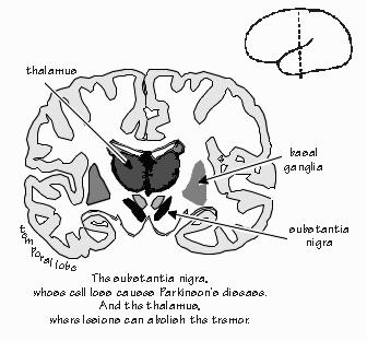 We talked about another brain disease before the dissection, which was Parkinson's Disease. I showed the students what part of the brain is affected when you have PD, which a small area called the substantia nigra. The reason it is called that is because it is one of the only naturally pigmented areas of the brain, and it looks like little iron filings scattered in a mustache shape deep in the brain. People don't have any symptoms until about 90% of the neurons there have died. They first notice a little twitch, like in their pinky finger, and then they gradually develop a tremor. The tremor can be treated for a while with various drugs that make it go away for a few hours, but then it keeps coming back and getting worse and worse. Eventually, the person has more and more problems and passes away, typically after 20 or more years of symptoms. For the homework, we will explore a surgical technique to help with PD symptoms.
The above image is a view of the brain by a coronal section, as illustrated in the small figure. We talked about cutting a brain horizontally and sagitally too. Here is a picture of the different ways of cutting the brain. During most of my research, I have done sagittal sections of brain, and that is what we did for the sheep brain dissection. To do a sagittal section, we cut the brain down the middle from above.
For the dissection itself, we followed the student guides and also this procedure. Some things we talked about during the dissection:
 |
| A view of the dissected sheep brain |
- Meningitis is when the membrane covering the brain (called the meninges) gets infected. It is very serious and can kill you, because swelling within the skull causes high pressure, which then squishes the brain and causes irreversible damage.
- The bulges all over the surface of the brain are called gyri (plural of gyrus), and the grooves are called sulci (plural of sulcus).
- There are a lot of nuclei in the brain (plural of nucleus), which are groups of neurons that do some specific function.
- The hypothalamus is a small structure near the brainstem that has several nuclei. One of them controls things like hunger, thirst, and sleep.
- The pituitary is a gland that releases hormones, including growth hormone to tell the body when it should grow.
- Sheep have large olfactory bulbs that they use for understanding smells in their world. Most mammals rely more on smell than we humans do, and their brain olfactory areas are proportionately larger.
- The cerebellum is important for learning motor skills, like riding a bike, mirror writing, and throwing a ball.
- My neuroscience program has several sets of prism glasses they use for demonstrations. You throw a ball into a basket very easily, then put on the glasses. Now, you start missing the basket. If you keep trying, your cerebellum helps make the right adjustments to your throwing and you start to get baskets again. Then when you take them off, you start missing again and need to keep practicing to adjust back to how to throw. This all happens within seconds to minutes! Learning to ride a bike takes a bit longer, but once you get the hang of it, you don't use your cerebellum much for that skill anymore.

- A man who had a major memory impairment due to stroke lost his ability to make new conscious memories. Everyone he met after his stroke he promptly forgot who they were. But he worked with researchers over many years who taught him motor skills like mirror writing. While he had no memory of ever trying the task, his skill improved at it, showing that the motor learning happened in a different part of the brain than his stroke damaged area.
- The corpus callosum is where axons from neurons in the brain cross over from one side to the other. Surprisingly, people function pretty well if their corpus callosum is cut or never forms.
- Ventricles in the brain are areas with fluid in them instead of cells. The fluid that the brain and spinal cord sit in is called CSF, or cerebrospinal fluid, and it helps keep the brain healthy.
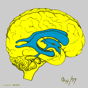 |
| The ventricles of the human brain |
No comments:
Post a Comment