Monday, April 29, 2013

Yummy Neurons!

This week, we made models of neurons -- out of candy!  It was a team effort, and it seemed like everyone was able to leave with a sugar high!  There was enough candy for all the parts of interest, and we even had some choices to make:

  • The cell bodies were the cupcakes.  Some cut off the tops and dug out the middle, and some just dug into the top of the cupcake to create space for the insides of our neurons.
  • Once we had space, in went the cytoplasm -- green Jell-O!
  • We each chose a nucleus for our cells -- either a large jelly bean or a cherry sour
  • Before putting the nucleus into the cytoplasm, we wrapped bits of endoplasmic reticulum around them -- fruit roll-ups
  • On some parts of our endoplasmic reticulum, we sprinkled some ribosomes, which makes it rough endoplasmic reticulum (smooth endoplasmic reticulum doesn't have ribosomes).  Most of us used red sprinkles for the ribosomes, but we discussed using nerds.  Some sprinkled nerds into the cytoplasm to represent the ribosomes that float loosely separate from the endoplasmic reticulum.
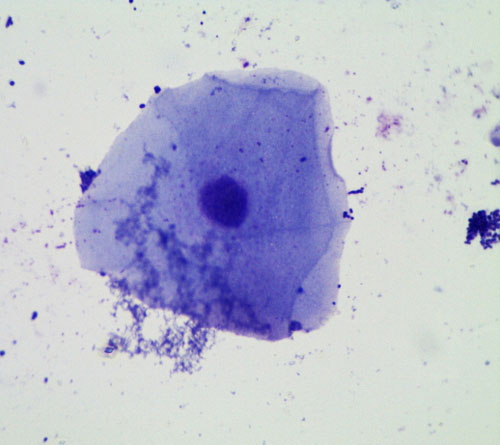
    Someone else's candy neurons; you can see organelles!
  • Next, we chose some mitochondria -- small jelly beans, about 3 per cell (most wanted the Starburst jelly beans, but a few used the Jelly Bellies)
  • Some of us added lysosomes to our cytoplasm -- nerds
  • One student who just got his braces put on used raisins for his organelles, since he couldn't have some of the gooier candies
  • Then, we added the key parts of neurons that make them special compared to other cells:
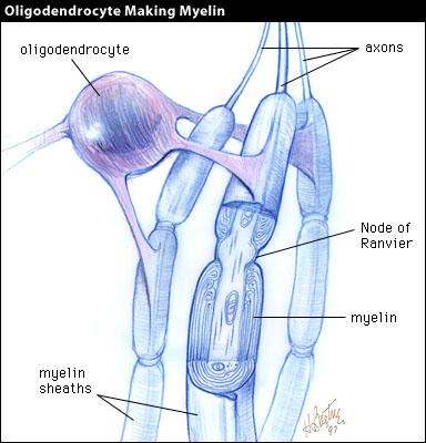
    • The axon was a Twizzler, and we wrapped it with myelin sheaths -- marshmallows
    • The dendrites were bits of sour licorice straws
    • We added tiny synaptic boutons, which are where the signal comes into the dendrites
    • To represent the neurotransmitter crossing the synapse to send the message from one cell to the next, we used the frosting
Finally, we created a large circuit with our neurons, by connecting them together.  We lined up our neurons and connected each axon to one of the next cell's dendrites (with neurotransmitter of course), and created a seven or eight cell circuit!  Most of our neurons did not survive long after that, moving bit by bit into our digestive systems.
Diagram of two neurons showing their synapse

To model how neuronal circuits work, we also lined up and held hands, to represent neurons connected together.  A message started at one end of the circuit and traveled to the other end.  The message was a hand squeeze, and each neuron had to send the message on to the next as soon as it was felt.  The last neuron said "Got it" when the message got through.  We all noticed that it was a bit slow the first time, but after repeating the process several times, the message passed very quickly.  Just like when the neurons get myelinated, so their messages travel much faster.

We send some more messages down our circuit using the game "Telephone" where one person whispers a message to the next person, who whispers it to the next.  One message sent that way arrived intact, but several others were changed or garbled.  The nervous system has many features to prevent that kind of thing from happening.  Its job is to send messages very quickly and very accurately all over our bodies.

Monday, April 15, 2013

Neuroscience intro

We are finally at my area of expertise -- neuroscience -- and we started by reading this page for homework and having a quiz show based on it.  Here were some of the main facts we went over:

  1. Brain cells or nerve cells are called neurons.
  2. Neurons have a nucleus, cytoplasm, mitochondria, and other organelles that are typical of animal cells.
  3. Neurons communicate with each other.
  4. The way they connect and communicate is through structures called synapses.
  5. Synapses are places where neurons communicate, but there is a teeny tiny gap between the two cells.
  6. The brain has approximately 100 billion neurons, and approximately 1 quadrillion synapses.
  7. The adult human brain weighs about 3 pounds
  8. Neurons have a long, skinny part called the axon that sends the message to the next cell.
  9. If you laid all of an adult human's neurons end to end, it would be about 600 miles.
  10. The brain is about 2% of the total body weight for an adult.
  11. Most neuroscientists have had 12 years of grade school, 4 years of college, and 4-7 years of graduate school.
We also discussed the insulation around neurons.  You can see in the image above that there are rod-like things going down the axon. That is actually from cells called oligodendrocytes that are near neurons that wrap themselves around the neurons to insulate them, like we use rubber to insulate wires.  The insulation makes the message travel faster, and as our brains get more insulation around their neurons, we become more able to do things.  Babies have hardly any of it, and new skills develop as their brains
develop in this way.  If you lose the insulation around your neurons, you become disabled in various ways because the brain doesn't function as well.
The insulation is very fatty, and because of that, areas of the brain and spinal cord with a lot of insulation look white.  Areas that mostly have the cell bodies of the neurons look gray.  That's why these areas are called white matter and gray matter -- they really look white and gray when you dissect them, as we will see.

We took a look at our sheep brains and saw that they have a thin
What our sheep brains look like
membrane over them.  This is not much real protection compared to the skull, but it helps regulate the fluid pressure inside the brain, which is very important.  Too much or too little pressure inside the brain can be a disaster!  We also saw the brainstem at the base of the brain, which is a huge bundle of mostly axons that are going to the spinal cord.  We noticed that the surface feels smooth, but you can see all sorts of wrinkles and twists like a small intestine.  We will discuss why that is and what it means.  Lastly, we weighed our brains, and they weighed about 150g.  Not very much!  They are very small compared with human brains!

Thursday, April 11, 2013

Awesome cell stuff!

In class i learned that the nucleus holds the hole blueprint to make a hole new cell!
And that the cell wall holds up the side of the plant cells!
It was really fun to look at the dyed slide!


A dyed slide!     

Monday, April 8, 2013

Slide-Making and Cells

This blog post covers what we've done for the past two class meetings.  Our overall focus for these two classes has been making slides, and for the second class, we discussed what cells are and the parts of cells.

Histology is the microscopic study of the cells and tissues of plants and animals.  Basically, that means that people put samples onto microscope slides and the look at them under the microscope to see what they can find out about the sample.  Most of the time, they add a stain to the sample, because you can't see very much without a stain.  Stains are usually dyes, and they are added to the sample because they often stick more to some parts of the sample than others, which lets you know things like where the edges of the cells are and where parts within the cells are.

Here is a picture of kidney tissue stained with H&E, which makes some parts of the cells pink, some red, and some dark purple.  It's a very common stain for human or animal tissues.
At some point, we would like to stain the tissues we are examining from our pigs with eosin dye, but it might not work very well for several reasons.  First, there is a complex staining process that will be hard for us to replicate, and second, we don't have the equipment to cut our samples super thin.  Most histology samples are about 4 microns thick, cut using a microtome.  A microtome works basically like a meat slicer and cuts very thin sections of your sample, that you then put onto a slide, stain, and examine under a microscope.  I will look into the possibility of us getting a tour of a histology facility on campus, as there are several decent ones in the medical school and vet school.

During the first class, we took samples of our own cheek cells and looked at them under a microscope.  Here's what they looked like without any stain on them and with methylene blue stain on them:
You can see that the blue stain lets us see the cells better, including the little oval in the center, which is the nucleus.


We also tried a purple stain called crystal violet, and cheek cells stained with it look like this: 

I got this image from the internet, and I think the stain makes bacteria dark purple, which would be the little tiny dots all over the place.  We have a lot of bacteria in our mouths, and this picture shows how much more bacteria we have than even our own skin cells.

We tried Eosin too and didn't see much, but also our microscope is dying.  The update is that the nosepiece, which is the part that has different magnification objectives, came off.  I don't think I can fix it, so I think we have to let that really cool microscope rest in peace.  I still have the dissecting microscope, and will look into getting another high magnification microscope.  

This past week, we discussed the parts of plant and animal cells, so that we can better understand what we're seeing when we look at cells under a microscope.  We assigned each student a cell part to represent, and discussed what each part does.  

First, Henry was the cell wall.  The cell wall is only found in plant cells, and it is a rigid structure that helps the plant stay upright, grow tall, and keep the cell protected.  Animal cells don't have one because they need to be more flexible, such as muscle cells that need to contract.

Then, Aaron was cell membrane.  Animal cells have a cell membrane to separate them from their environment, and so do plant cells.  It is more flexible than the cell wall.

Elinor was the nucleus, which is a very important organelle.  (Organelles are the small parts inside cells that do things for the cell, like organs in our bodies do things for our entire body).  The nucleus contains the DNA, which is the complete blueprint for making the whole organism.  It's like a recipe book, and each recipe makes a protein.  If you follow all the recipes in the right order, you make an Elinor or a frog, whatever the blueprint (DNA) says.  Every cell in our bodies contains a complete blueprint for making us, with two exceptions.  Red blood cells are born with nuclei (plural of nucleus), but after a few weeks, once they have made the proteins they will need to carry oxygen around the body, they spit out their nuclei and live for another month with no nucleus.  Sex cells (the sperm and the egg) each only have half of the blueprint, such that when they combine, a complete blueprint for a person is made.  If each had a complete set of DNA, there would be too much and an organism can't be made if a cell has a double blueprint (too much DNA).

Jett was the vacuole, which is the large white space we can see in the cells.  In plant cells, there is a large, central vacuole that takes up most of the space in the cell, but in animal cells, if they even have vacuoles, they are small and don't take up much space.  It's not clear to me exactly what they do.  

Anne Sophie was the endoplasmic reticulum, which will always be found around the nucleus.  It is a stack of tubes that take information from the DNA blueprint to turn into proteins.

Esme was the lysosome, which is a small blob in which proteins that are no longer needed get broken down.

Ben was the mitochondria, which makes the energy for the cell.  Plants have chloroplasts in some of their cells, which take energy from the sun to start making sugar.  All cells have mitochondria that take sugars from photosynthesis or from eating foods and then convert them into energy the cell can use to make things and live.

Our plant cell
Jordan was the cytoplasm.  All the organelles float in a liquid called the cytoplasm.

Liam was the ribosomes, and some ribosomes just float around on their own while many of them are actually in the endoplasmic reticulum.

There are more organelles than we discussed, but we're going to leave it at that for now.

Next, we pulled off the skin layer of some onion bulbs and looked at them under the microscope.  Here are some pictures we took.
High magnification view of our onion cells
Unstained by slightly dried out onion skin

Onion cells stained with Iodine-propidium iodide
 
Lastly, we looked at a section of lilac leaf that was stained to show us the different cells.