Histology is the microscopic study of the cells and tissues of plants and animals. Basically, that means that people put samples onto microscope slides and the look at them under the microscope to see what they can find out about the sample. Most of the time, they add a stain to the sample, because you can't see very much without a stain. Stains are usually dyes, and they are added to the sample because they often stick more to some parts of the sample than others, which lets you know things like where the edges of the cells are and where parts within the cells are.
 |
| Here is a picture of kidney tissue stained with H&E, which makes some parts of the cells pink, some red, and some dark purple. It's a very common stain for human or animal tissues. |
During the first class, we took samples of our own cheek cells and looked at them under a microscope. Here's what they looked like without any stain on them and with methylene blue stain on them:


You can see that the blue stain lets us see the cells better, including the little oval in the center, which is the nucleus.
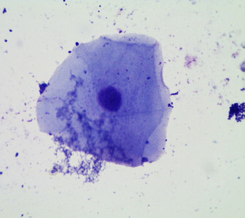 We also tried a purple stain called crystal violet, and cheek cells stained with it look like this:
I got this image from the internet, and I think the stain makes bacteria dark purple, which would be the little tiny dots all over the place. We have a lot of bacteria in our mouths, and this picture shows how much more bacteria we have than even our own skin cells.
We tried Eosin too and didn't see much, but also our microscope is dying. The update is that the nosepiece, which is the part that has different magnification objectives, came off. I don't think I can fix it, so I think we have to let that really cool microscope rest in peace. I still have the dissecting microscope, and will look into getting another high magnification microscope.
First, Henry was the cell wall. The cell wall is only found in plant cells, and it is a rigid structure that helps the plant stay upright, grow tall, and keep the cell protected. Animal cells don't have one because they need to be more flexible, such as muscle cells that need to contract.
Then, Aaron was cell membrane. Animal cells have a cell membrane to separate them from their environment, and so do plant cells. It is more flexible than the cell wall.
Elinor was the nucleus, which is a very important organelle. (Organelles are the small parts inside cells that do things for the cell, like organs in our bodies do things for our entire body). The nucleus contains the DNA, which is the complete blueprint for making the whole organism. It's like a recipe book, and each recipe makes a protein. If you follow all the recipes in the right order, you make an Elinor or a frog, whatever the blueprint (DNA) says. Every cell in our bodies contains a complete blueprint for making us, with two exceptions. Red blood cells are born with nuclei (plural of nucleus), but after a few weeks, once they have made the proteins they will need to carry oxygen around the body, they spit out their nuclei and live for another month with no nucleus. Sex cells (the sperm and the egg) each only have half of the blueprint, such that when they combine, a complete blueprint for a person is made. If each had a complete set of DNA, there would be too much and an organism can't be made if a cell has a double blueprint (too much DNA).

Jett was the vacuole, which is the large white space we can see in the cells. In plant cells, there is a large, central vacuole that takes up most of the space in the cell, but in animal cells, if they even have vacuoles, they are small and don't take up much space. It's not clear to me exactly what they do.
Anne Sophie was the endoplasmic reticulum, which will always be found around the nucleus. It is a stack of tubes that take information from the DNA blueprint to turn into proteins.
Esme was the lysosome, which is a small blob in which proteins that are no longer needed get broken down.
Ben was the mitochondria, which makes the energy for the cell. Plants have chloroplasts in some of their cells, which take energy from the sun to start making sugar. All cells have mitochondria that take sugars from photosynthesis or from eating foods and then convert them into energy the cell can use to make things and live.
 |
| Our plant cell |
Liam was the ribosomes, and some ribosomes just float around on their own while many of them are actually in the endoplasmic reticulum.
There are more organelles than we discussed, but we're going to leave it at that for now.
Next, we pulled off the skin layer of some onion bulbs and looked at them under the microscope. Here are some pictures we took.


Lastly, we looked at a section of lilac leaf that was stained to show us the different cells.
 |
| High magnification view of our onion cells |
 |
| Unstained by slightly dried out onion skin |
 |
| Onion cells stained with Iodine-propidium iodide |
Lastly, we looked at a section of lilac leaf that was stained to show us the different cells.
No comments:
Post a Comment